
Fig. 1.
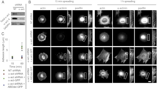
Binding of α-actinin to actin enables adhesion maturation. (A) Western blot showing levels of total α-actinin and α-actinin 4 in cells transfected with a shRNA with a NT or α-actinin 4 targeting (α-act) sequence. (B) Cells transfected with NT or α-actinin shRNA and rescued with FL or ABDdel α-actinin-GFP stained after 15 min or 1 h for F-actin, α-actinin, and paxillin. In rescue cells, α-actinin image is the corresponding GFP construct. Expanded views amplify the areas marked with a white rectangle. (Scale bar, 20 µm.) (C) Quantification of paxillin adhesion length for the different conditions at the 15-min and 1-h time points. Both NT-transfected cells and α-actinin depleted cells rescued with FL α-actinin-GFP experienced significant increases in adhesion size with time (P < 0.01), whereas α-actinin depleted cells either not rescued or rescued with ABDdel-GFP did not (n ≥ 6 cells measured on 2 different days).