
Fig. 6.
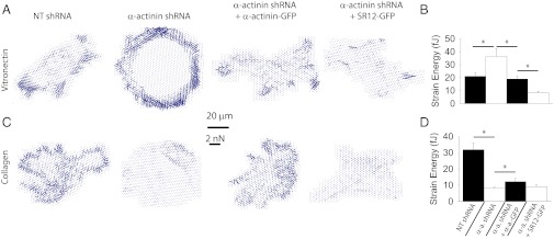
Role of α-actinin in cell contractility depends on matrix coating. (A) Vector plots with arrows depicting the magnitude and direction of forces exerted on 1-µm pillar arrays coated with vitronectin. Cells were transfected with either NT or α-actinin shRNA and rescued with FL or SR12 α-actinin-GFP. (Scale bar, 20 µm; force scale bar indicates the length of a force arrow of 2 nN.) (B) Corresponding quantification of average forces (strain energy) exerted by cells. (C and D) Same results on collagen I-coated pillars. *P < 0.05, n ≥ 11 cells measured on 2 different days.