Fig. 4.
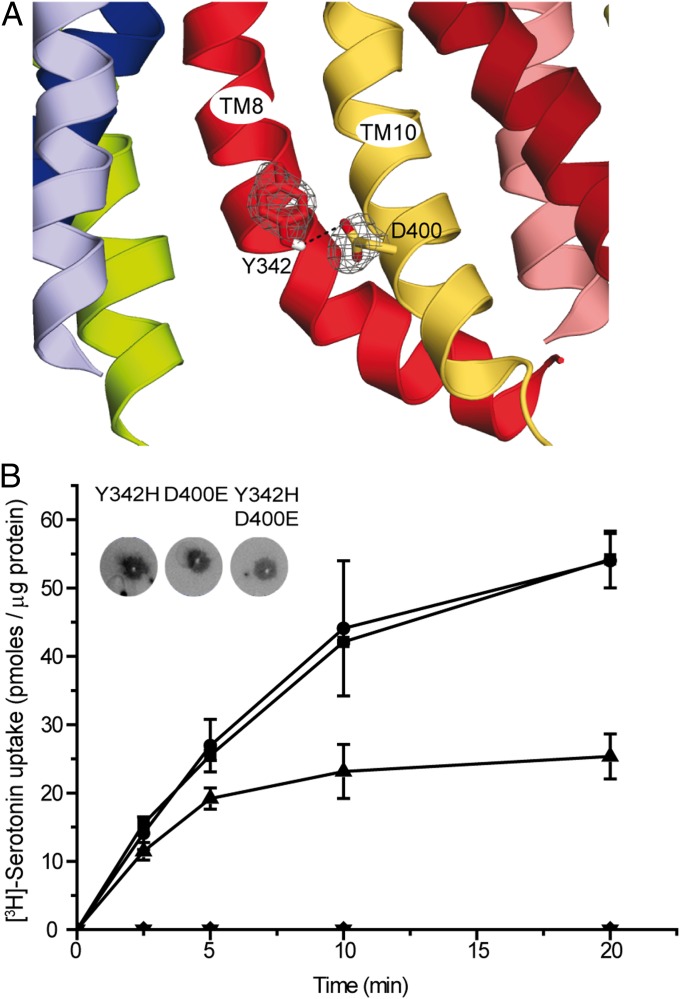
Predicted interaction between residues Y342 and D400 in TMs 8 and 10. (A) The model of rVMAT2 in a cytoplasm-facing state is viewed along the plane of the membrane focusing on the cytoplasmic half of the protein, which is oriented with the cytoplasm toward the bottom. Side chains of Y342 and D400 are shown as sticks, and the probability density of their positions in the 100 top models is shown as a gray mesh. (B) [3H]-serotonin uptake. Liposomes (1 µL) reconstituted with rVMAT2 (■) D400E (▲), Y342H (●),Y342H-D400E (▼), and vector with no gene (mock) (◆) were assayed for serotonin transport as described in Materials and Methods. (Inset) Quantitation of protein amounts in proteoliposomes using a dot blot assay. Results presented are from duplicate experiments; error bars indicate SE. Experiments were repeated at least twice.

