Fig. 6.
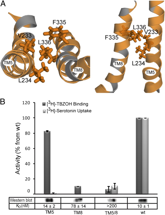
Predicted hydrophobic interactions between TM5 and TM8. (A) Close-up of TMs 5 and 8 in the cytoplasm-facing model of rVMAT2 viewed as in Fig. 2A (Left) and along the plane of the membrane (Right) with the cytoplasm toward the bottom. Hydrophobic residues (Ala, Gly, Val, Ile, Leu, Phe, Met, and Trp) are colored orange. Side chains of L336, F335, L234, and V233 are shown as sticks. (B) [3H]-TBZOH binding to HEK239 permeabilized cells, [3H]-serotonin transport for V233A/L234A (TM5), F335A/L336 (TM8), and the quadruple mutant V233A/L234A/F335A/L336 (TM5/8), and Western blot relative to wild-type rVMAT2. Results presented are from duplicate experiments; error bars indicate SE. Experiments were repeated at least twice.

