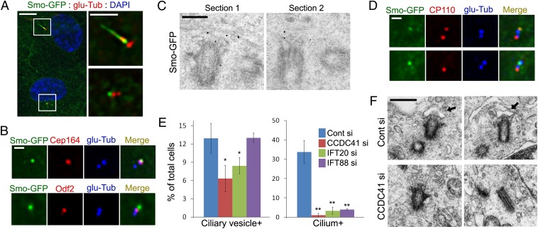
Fig. 5.
CCDC41 may be involved in the docking of the primary ciliary vesicle. (A) RPE1 cells stably expressing Smo-GFP (RPE1-SmoGFP) were serum-starved for 16 h and stained with anti–glu-Tub antibody. (Right) Higher-magnification view of the boxed areas at Left. (B) RPE1-SmoGFP cells were double-stained with the indicated antibodies. (C) PRE1-SmoGFP cells were serum-starved for 16 h, and processed for immuno-EM with anti-EGFP antibody. (D) RPE1-SmoGFP cells were serum starved (Upper, 8 h; Lower, 16 h), and double stained with anti-CP110 and anti–glu-Tub antibodies. (E) RPE1-SmoGFP cells were transfected with the indicated siRNAs for 3 d (cells were serum-starved for the final 16 h) and stained with anti–glu-Tub antibody. Error bars represent SD (n = 3 independent experiments; *P < 0.05 and **P < 0.01, t test). (F) RPE1 cells were transfected with siRNAs for 3 d (cells were serum-starved for the final 16 h) and processed for transmission EM analysis. Arrows indicate vesicles anchored to the distal end of the centriole. (Scale bars: A, Left, 10 μm; A, Right, 5 μm; B and D, 1 μm; C, 200 nm; F, 500 nm.)

