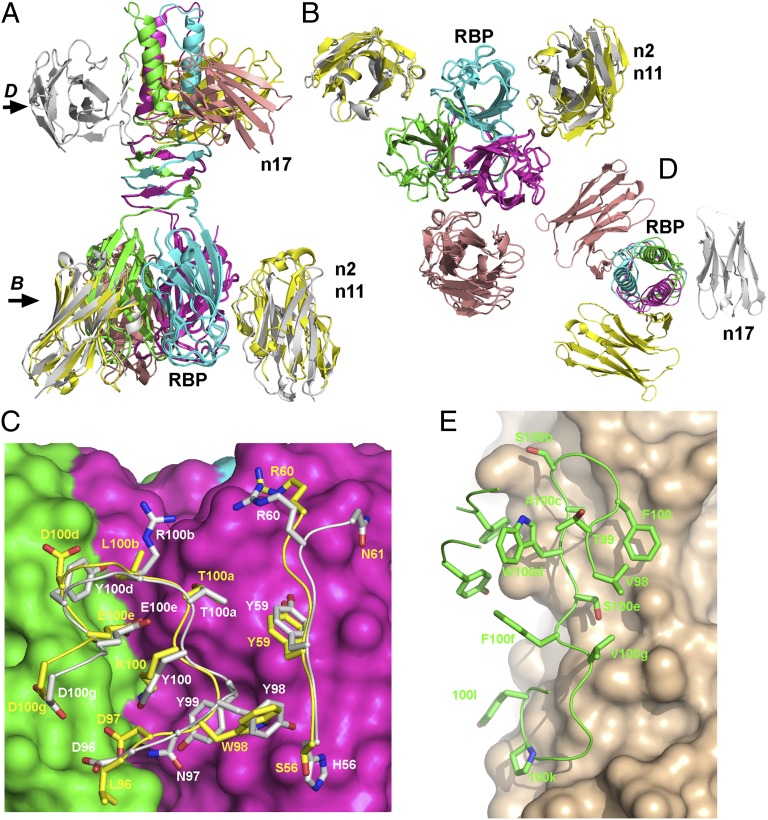
Fig. 6.
X-ray structures of the various complexes formed by BppL and nanobodies 2, 11, and 17. (A) Ribbon view of the superposition of the three structures of the complexes; (B) 90° view (compared with A) of the BppL complexes with nanobodies 2 and 11 sliced at the level of the head domains. (C) Detailed view of the interaction area between BppL head domains (pink and green surfaces) and CDR2 and -3 of nanobodies 2 (white sticks) and 11 (yellow sticks); (D) 90° view (compared with A) of the BppL complexes with nanobody 17 sliced at the level of the stem domains. (E) Detailed view of the interaction area between BppL stem and β-helix domains (beige surface) and CDR3 of nanobody 17 (green sticks). In C and E, numbering is according to the work by Kabat et al. (47)

