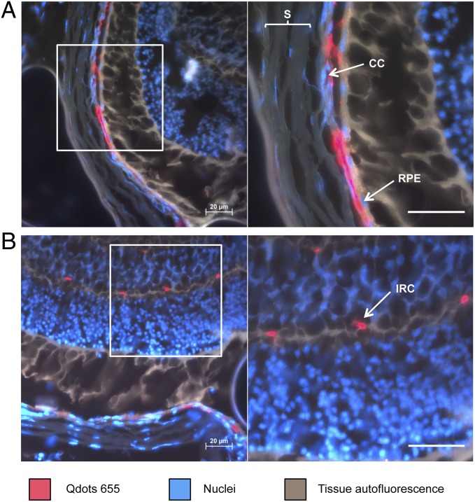
Fig. 4.
Investigation of cyclo(RGDfC)-Qdot location in the posterior segment of the eye. The major part of Qdots is clearly located associated to the endothelia in the choriocapillaris (A). Further accumulation of cyclo(RGDfC)-Qdots was detectable in intraretinal capillaries (B). CC, choriocapillaris; IRC, intraretinal capillaries; RPE, retinal pigment epithelium; S, sclera. (Scale bars: 20 µm.)

