
Figure 2. Actin cytoskeleton organization on different hydrogels.
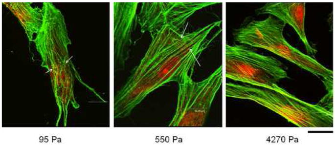
Immunofluorescent staining with Alexa Flour 488-Phalloidin shows that actin fibers become more stretched and organized with increasing substrate mechanics. Arrows indicate areas of filament discontinuity and kinks/bucklings. Scale bar = 16 μm.