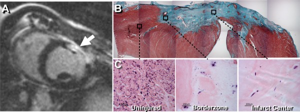
Fig. 1.

(a) Contrast-enhanced MRI confirming the presence of infarcted myocardium (arrow) at 3 weeks post cryoinjury. (b) Masson's trichrome stain of the 30 day old myocardial infarct site showing full transmural infarct. (c) Endogenous alkaline phosphatase staining, with eosin counterstain, showing endothelial cells within the uninjured myocardium, infarct border zone, and infarct center.
