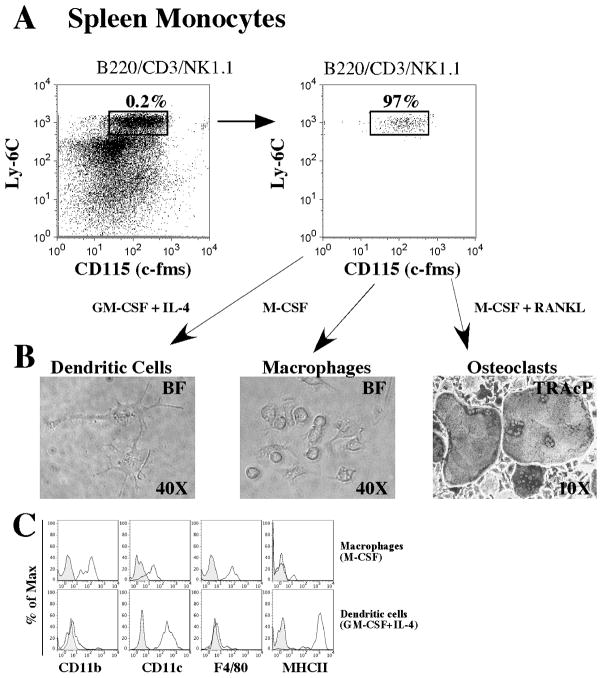
Figure 5. Evidence of a common splenic monocyte progenitor for osteoclast, macrophages and dendritic cells in vitro.
A) Cells with the phenotype LYM− CD11b+ Ly6C+ CD115+ were sorted to homogeneity and plated at a density of 5,000 cells in GM-CSF (30ng/mL) plus IL-4 (30ng/mL) for 8 days, M-CSF (30ng/mL) for 8 days or M-CSF (30ng/mL) plus RANKL (30ng/mL) for 8 days in 96 well plates. B) Images of cells developed at the end of the culture period. DC- and macrophage-like cells are visualized by phase contrast (right and middle) and osteoclasts are visualized by light microscopy after TRAcP staining (right). C) After culture, cells were detached from 96 well plates and analyzed for their cell surface expression of signature cell surface markers for DC and macrophages. Cells cultured with GM-CSF plus IL-4 (top row, histograms) show preferential expression of cell surface molecules related to DCs (CD11c+MHCII+F4/80−CD11blow), and cells cultured with M-CSF (bottom row) show preferential expression of cell surface markers related to macrophages (CD11b+F4/80+CD11clowMHCII−).

