Fig. 2.

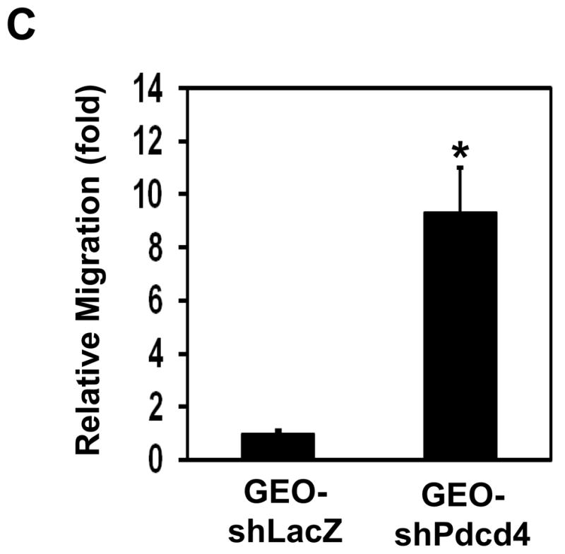
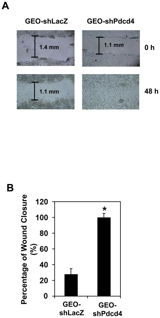
Knockdown of Pdcd4 promotes cell migration. (A) Wound-healing assays. GEO-shLacZ and GEO-shPdcd4 cells were seeded onto a 24-well plate. At 100% confluence, cell monolayer was scratched with a plastic tip. At 0 h and 48 h after the scratch, the distance of wounded monolayer was measured under a microscope equipped with a digital camera. The representative images are shown. (B) The percentage of wound closure. Each value is expressed as the mean ± standard deviation (SD) of three independent experiments. The asterisk indicates significant difference compared with GEO-shLacZ cells as determined by one-way ANOVA (P<0.01). (C) Boyden chamber migration assays. The number of cells that has migrated to the lower side of membrane surface was determined by counting 7 areas. The migration capacity of GEO-shLacZ cells is designed as 1. Two independent experiments were performed with triplicates for each sample. The representative data are shown and expressed as the mean ± SD. The asterisk indicates a significant difference compared with GEO-shLacZ cells as determined by one-way ANOVA (P<0.05).

