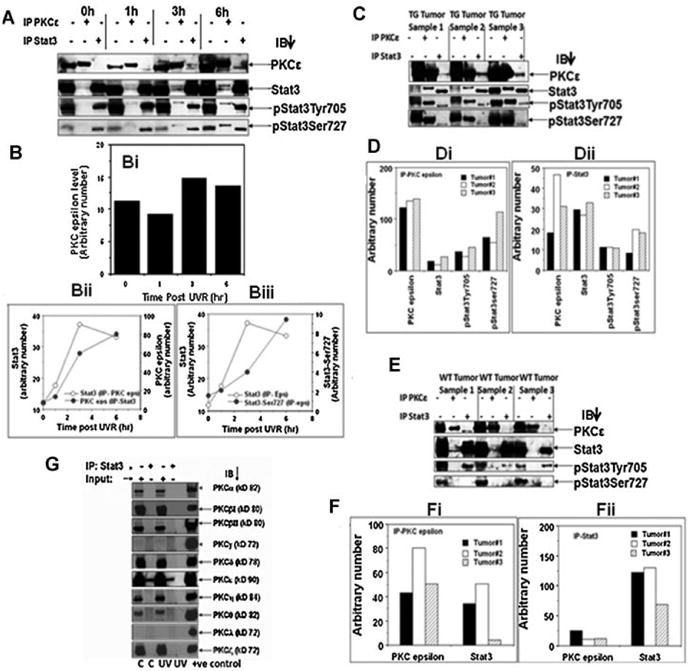
Figure 2.

UVR-dependency and isoform specificity of PKCε– Stat3 interaction. A: PKCε transgenic mice (line 224) were exposed once to 2 kJ/m2 UVR and sacrificed 1, 3, and 6 h after UVR exposure (3 mice/group). Epidermal lysates were prepared as described in Materials and Methods Section. Epidermal lysates from three mice were pooled. Epidermal lysate (100 μg) was immunoprecipitated with either PKCε or Stat3 and immunoblotted with the indicated antibodies. B: Quantification of the Western blots shown in (A). Bi: Effect of UVR treatment on basal level of PKCε expression (input), Bii–iii: PKCε–Stat3 interaction increases following UVR treatment. C,E: PKCε transgenic mice (TG) (line 224) and wildtype littermate (WT) were exposed thrice weekly to UVR (1 kJ/m2) for development of SCC, 23 wk. Mice were then sacrificed and the SCC (TG Tumor, WT Tumor) was excised and total epidermal lysates were prepared. Total epidermal cell lysate (100 μg) was immunoprecipitated using Stat3 or PKCε antibody. Immunoprecipitated proteins were then subjected to SDS–PAGE and blotted with PKCε, Stat3, pStat3Tyr705, and pStat3Ser727. D,F: Quantification of blots shown in C (TG Tumor) and E (WT Tumor). Di: ip PKCε, (Dii) ip Stat3, (Fi) ip PKCε, (Fii) ip Stat3. G: PKCε transgenic mice (line 215) were exposed to UVR (2 kJ/m2) four times (Monday, Wednesday, Friday, and Monday). The mice were then sacrificed 3 h post-last UVR exposure. Total epidermal cell lysate (100 μg) was immunoprecipitated using Stat3 antibody. Stat3-immunoprecipitated proteins were subjected to SDS–PAGE and immunoblotted with the indicated PKC isoform (α, β, δ, ε, γ, η, ι, λ) antibody. Input is 25 μg of epidermal lysate. Positive controls for PKC isoforms as described in Figure 1. C: control (untreated) mice, UV: 3 h post-final UVR exposed mice.
