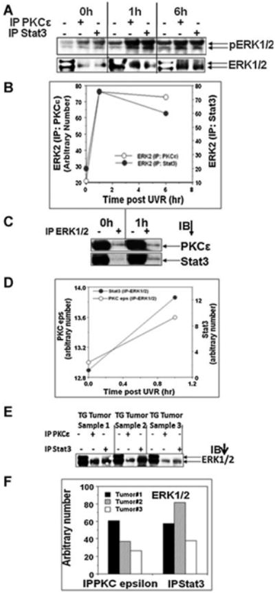
Figure 3.

PKCε and Stat3 co-immunoprecipitate with ERK1/2. Same epidermal lysate, prepared for experiment described in Figure 2, was used. A: Epidermal lysate (100 μg) was immunoprecipitated with either PKCε or Stat3 and immunoblotted with phospho and total ERK1/2 antibodies. Input was 25 μg epidermal protein. B: Quantification of the Western blot in (A). C: Epidermal lysate (100 μg) was immunoprecipitated with ERK1/2 and immunoblotted with PKCε and Stat3. Input was 25 μg epidermal protein. D: Quantification of the Western blot in C. E: PKCε line 224 and wildtype littermates were exposed thrice weekly to UVR (1 kJ/m2) for 23 wk. Mice were then sacrificed and the SCC was excised and total epidermal lysate was prepared. Total epidermal cell lysate (100 μg) was immunoprecipitated using Stat3 or PKCε antibody and blotted for ERK1/2. Input was 25 μg epidermal protein. F: Quantification of the Western blot in (E).
