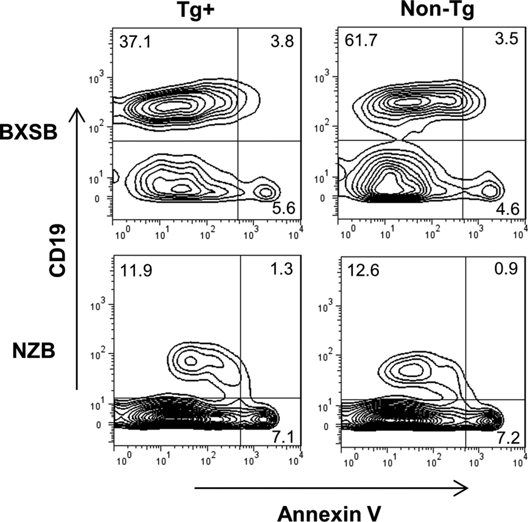
Figure 8.
Apoptosis in BXSB and NZB splenic lymphocytes. RBC-depleted splenocytes were stained with annexin V, 7-AAD, and CD19 and flow cytometric data acquired within 30 minutes of staining. Shown are representative plots gated initially on lymphocytes by light scatter properties. The % of lymphocytes are indicated in the plots; the percent annexin+ of B cells in these representative plots are 9.3% Tg+ and 5.4% non-Tg for the BXSB, and 9.8% Tg+ and 6.7% non-Tg for the NZB.

