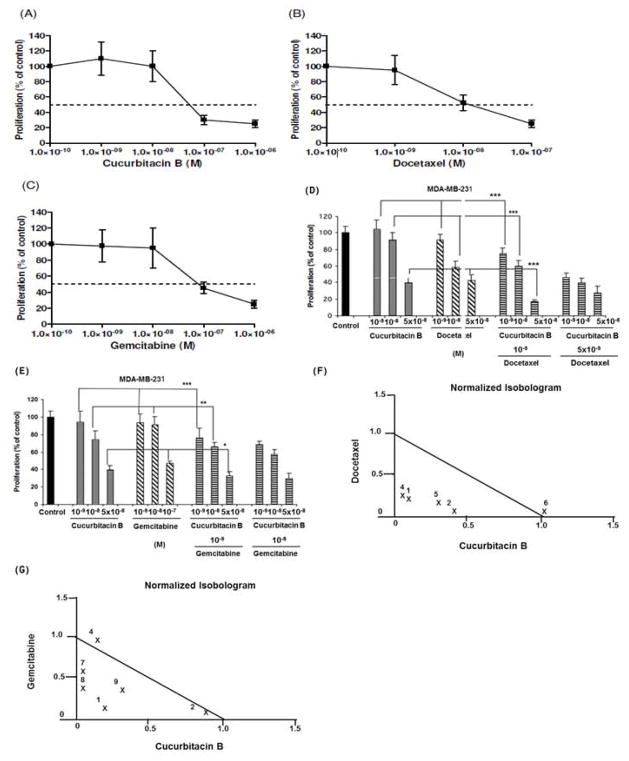
Figure 1. Effect of CuB, DOC and GEM on proliferation of MDA-MB-231 cells in vitro.
MTT-proliferation assays of MDA-MB-231 cells treated for 96 hrs with different concentrations of the following drugs: Panels (A) CuB, (B) DOC, (C) GEM, (D) CuB/DOC or (E) CuB/GEM. (F, G) Normalized isobolograms. Numbers represent different concentrations of Panel (F) CuB/DOC, Panel (G) CuB/GEM. Panel (F) #1: DOC 10−9 mol/L+ CuB 10−9 mol/L; #2: DOC 10−9 mol/L + CuB 10−8 mol/L; #4: DOC 5 × 10⁻⁹ mol/L + CuB 10−9 mol/L; #5: DOC 5 × 10⁻⁹ mol/L + CuB 10−8 mol/L; #6: DOC 10−8 mol/L + CuB 5 x 10⁻⁸ mol/L. Panel (G) #1: GEM 10−9 mol/L + CuB 10−9 mol/L; #2: GEM 10−9 mol/L + CuB 10−8 mol/L; #4: GEM 10−8 mol/L + CuB 10−9 mol/L; #7: GEM 10−7 mol/L + CuB 10−9 mol/L; #8: GEM 10−7 mol/L + CuB 10−8 mol/L; #9: GEM 10−7 mol/L + CuB 5 x 10⁻⁹ mol/L; *: p < 0.05; **: p < 0.01; ***: p< 0.001.

