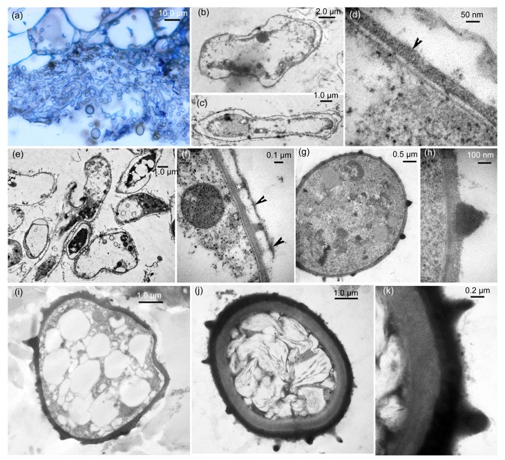
Fig. 2.
Development of teliospore walls as seen by light microscopy and transmission electron micrograph (TEM)
(a) Light microscopy. Abundant hyphal segments produced from sporogenous hyphae in a large intercellular space. (b–k) TEM. (b, c) Plasmolyzed process of hyphal segments: (b) a hyphal segment being plasmolyzed; (c) a completely plasmolyzed hyphal segment. (d) Exosporium (arrowhead) of a young teliospore produced on plasma membrane in plasmolyzed process. (e) Warty ornamentation formation and fungal cell walls being degraded. (f) A magnified view of warty ornamentation (*) with the remnant (arrowheads) of fungal cell wall in (e). (g) A regularly young teliospore with electron-dense warty ornamentation. (h) Magnified view of a conical wart in (g). (i) A young teliospore with electron-dense exosporium and spines. (j) A well-developed wall of teliospore with endosporium, exosporium, and warts. (k) Magnified view of a teliospore wall in (j)

