
Figure 1.
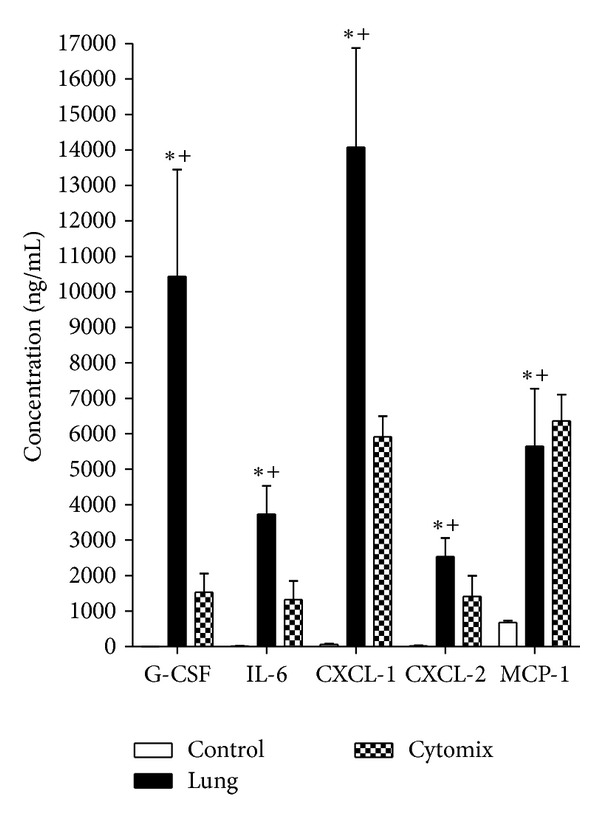
Absolute increase in selected cytokine concentrations in MLEC media. G-CSF, IL-6, CXCL-1, CXCL-2, and MCP-1 concentrations from MLECs exposed to control uncirculated perfusate (open bar), lung perfusate (solid bar), and cytomix (checkered bar). (*P < 0.05 concentration after 8 h incubation on MLEC versus concentration before incubation on MLEC (0 h), + P < 0.05 versus control perfusate after both were incubated on MLEC cultures for 8 h, ±SEM). Where open bars are not apparent (G-CSF, IL-6, CXCL-1, CXCL-2), the increase is too small to print.