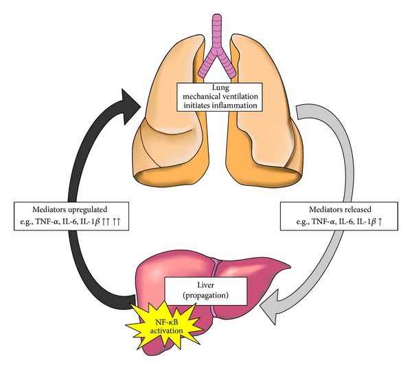
Figure 7.

Proposed inflammatory pathway diagram. Inflammation initiated in the lung releases inflammatory mediators (light grey arrow, right side) which then translocate to peripheral organs (e.g., liver). These organs amplify the inflammatory signal, through an NF-κB-dependent pathway, leading to further release of inflammatory mediators, which then travel back to the lung, and/or other peripheral organs (dark grey arrow) where the signal is further propagated in a feed-forward mechanism of acute inflammation.
