
Figure 6.
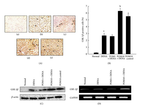
Effects of PE on GSK-3β expressions during DENA-evoked hepatic preneoplasia in rats. (A) Representative immunohistochemical localization of GSK-3β (magnification: 100x) in different rat groups. Several groups are (a) normal control; (b) DENA control; (c) PE 1 g/kg plus DENA; (d) PE 10 g/kg plus DENA; and (e) PE 10 g/kg control. (B) Quantification of hepatic GSK-3β-immunopositive cells. One thousand hepatocytes were counted per animal, and the results were based on 4 animals per group. Each bar represents the mean ± SEM (n = 4). a P < 0.001 as compared to normal group; b P < 0.001 as compared to DENA control. (C) Representative Western blot indicating protein expression levels of GSK-3β in various experimental groups and (D) representative RT-PCR analysis of GSK-3β expression in various groups of rats. Total hepatic RNA was isolated, subjected to reverse transcription, and resulting cDNA was subjected to RT-PCR analysis using specific primer sequence. The GAPDH was used as the housekeeping gene.