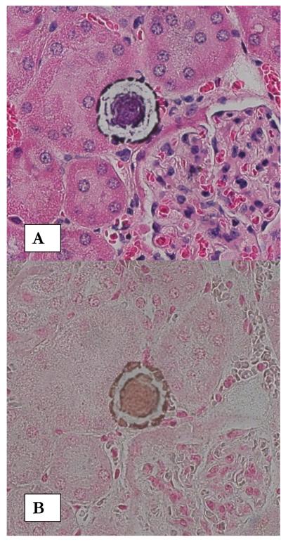
Figure1.
Calcium phosphate deposition in kidneys of NPT2a -/- mice. A. H&E stained paraffin section of a kidney from a 13 month old mouse showing what appear to be interstitial deposits in the renal cortex. Renal tubules are completely devoid of any crystals. Deposits show ring like substructure. Original magnification X 40. B. Von Kossa staining of a similar crystal deposit illustrates that it is calcium phosphate crystals.

