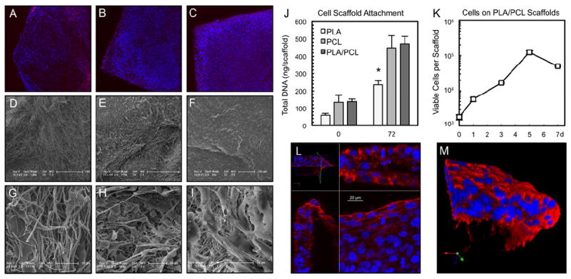
Fig. 2. Electrospun scaffolds support the growth of 3T3 mouse fibroblasts.

(A, B, C) Low magnification fluorescent microscopy. (D, E, F) Low and (G, H, I) high magnification SEM. In columns, from left to right are, PLA (A, D, G), PCL (B, E, H) and PLA/PCL composite (C, F, I) scaffolds. The cells form a confluent sheath, seen particularly well on PLA/PCL scaffolds. (J) After a 4-hour static seeding period, PCL and PLA/PCL scaffolds retain approximately twice as many cells as PLA scaffolds, a relationship that is statistically significant after a 72 hour in vitro culture period (* p=0.01, n≥4 per time point). (K) PLA/PCL scaffolds support long term in vitro culture and proliferation of cells (n=5 per time point). A decrease in viable cell numbers occurs by day 7 as the cells reach confluence on scaffolds with a finite surface area (2.25 cm2). (L) Confocal laser scanning microscopy stack showing cell nuclei growing in multiple layers in all three dimensions on PLA/PCL scaffolds. (M) Rendered reconstruction of cells growing in three dimensions on the PLA/PCL scaffold (edge shown for clarity). Actin in red, nuclei in blue.
