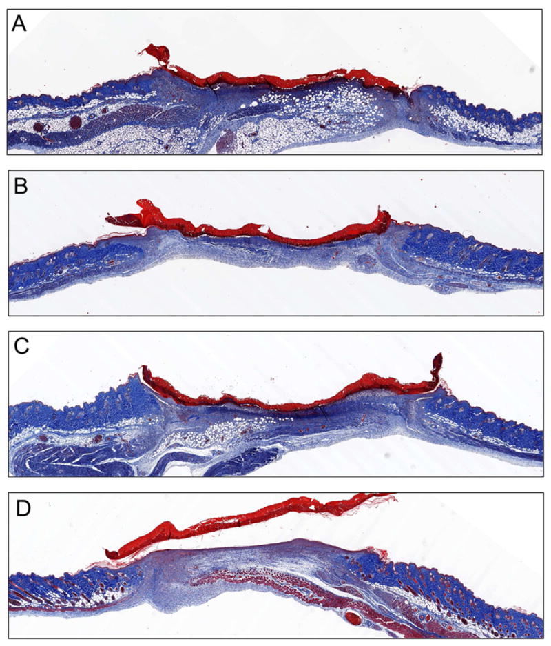
Fig. 6. Histological sections of wounds on day 5.

Direct comparison Masson’s trichrome stained sections of (A) control, (B) blank scaffold, (C) gWiz empty plasmid and (D) pKGF DNA scaffold treated wounds at day 5 reveals a much higher degree of re-epithelialization and structural maturity of the dermis in pKGF DNA scaffolds as compared to both other scaffolds and untreated wounds.
