
Fig. 2.
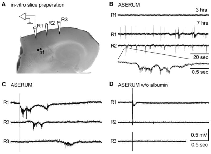
Spontaneous epileptiform activity after cortical exposure to serum components. a Photograph of the transverse brain slice preparation demonstrating the positioning of extracellular recording (R1–3) and stimulation (St) electrodes. b Representative traces of spontaneous activity recorded 3 and 7 h after exposure of the slice to artificial serum solution (ASERUM). Note the appearance of spontaneous hyper-synchronization of electrical activity within the cortex, as shown in simultaneous recordings from R1 and R2. c, d Representative electrophysiological traces demonstrating the response to brief electrical stimulation of afferent fibers in the white matter. Abnormally propagating epileptiform activity was evoked 5–7 h after incubation in ASERUM (c), but did not appear in slices incubated in albumin-free ASERUM (d) or ACSF (data not shown)