Fig. 2.
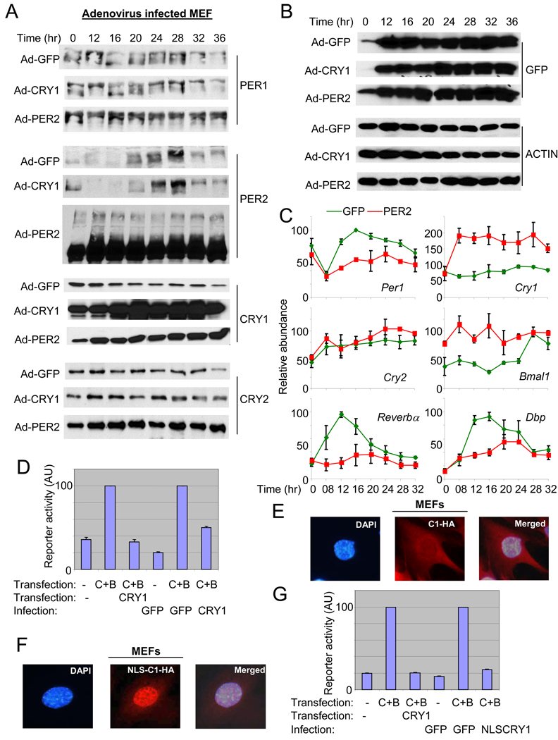
Constitutive expression of PER2, and not CRY1, disrupts PER protein rhythms and mRNA rhythms of clock and clock-controlled genes. (A) (B) MEFs were infected and serum shocked as described in Fig 1. Both endogenous (the fusion protein; top band) and exogenous (bottom band) PER2 are shown for PER2 immunoblot of PER2-MEFs. The adenoviral vector has dual CMV promoters, one default CMV promoter for GFP and the other one for the gene of interest (He et al., 1998), in our case PER2 or CRY1. Thus, similar intensities of GFP signal indicate similar viral titers among the three groups. (C) Quantitation of mRNA levels of clock and clock-controlled genes in Adenovirus GFP (green)- and Adenovirus PER2 (red)-MEFs. mRNA levels were measured by quantitative real time PCR. Data are shown as mean+/−SEM of three experiments. For some data points, error bars (if less than 3) are obscured by the square symbols. (D) The viral CRY1 can inhibit CLOCK:BMAL1-driven transcription in reporter assays. The inhibitory activity between plasmid (pcDNA3.1) and adenoviral CRY1 on CLOCK:BMAL1-driven transcription was compared in transient transfection assays. The first three reactions (bars) were done only with plasmid transfection but the other three reactions were performed with transfection (CLOCK and BMAL1) and viral infection (CRY). Results are shown as mean+/−SEM of triplicate samples and are representative of three independent experiments. (E) (F) The viral CRY1-HA was both cytoplasmic and nuclear (E), but the viral NLS-CRY1-HA was predominantly nuclear (F). The overexpressed viral CRY1-HA was visualized with immunofluorescence using anti-HA antibody. These single cells correspond to the cells in white boxes in Suppl Fig 2B–C, magnified as representatives. (G) The inhibitory activity of plasmid (pcDNA3.1)-expressed CRY and adenoviral NLS-CRY1 on CLOCK:BMAL1-driven transcription was compared in COS7 cells, as in (D).

