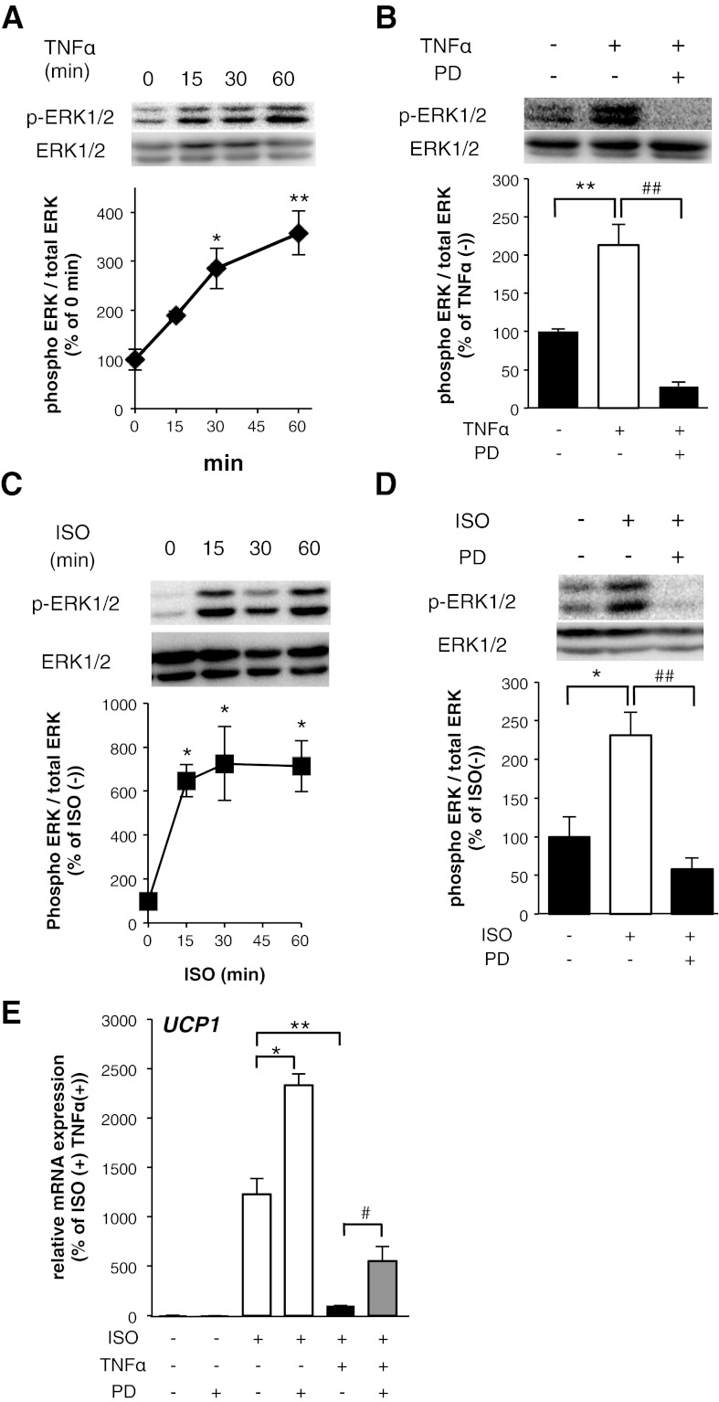
Fig. 5.
ERK activation induced by TNF-α mediated the suppression of UCP1 mRNA induction in 10T1/2 adipocytes. Total cell lysates (45 μg protein/lane) were examined by Western blot assay using the indicated antibodies. 10T1/2 adipocytes were exposed to TNF-α for the indicated times (A) and with PD98059 (PD) simultaneously for 15 min (B). 10T1/2 adipocytes were exposed to ISO for the indicated times (C) and with PD98059 simultaneously for 15 min (D). The band intensities were quantified using ImageJ software. The results were calculated using data of independent experiments repeated multiple times. *P < 0.05, **P < 0.01, compared with nonstimulated 10T1/2 adipocytes. ##P < 0.01, compared with TNF-α- or ISO-stimulated 10T1/2 adipocytes. E: mRNA expression levels of UCP1 in 10T1/2 adipocytes cultured with TNF-α with or without PD98059 for 12 h before ISO treatment (10 μM) for 8 h. Values are means ± SE for 3–4 wells. *P < 0.05, **P < 0.01, compared with ISO-stimulated 10T1/2 adipocytes. #P < 0.05, compared with ISO and TNF-α-stimulated 10T1/2 adipocytes.

