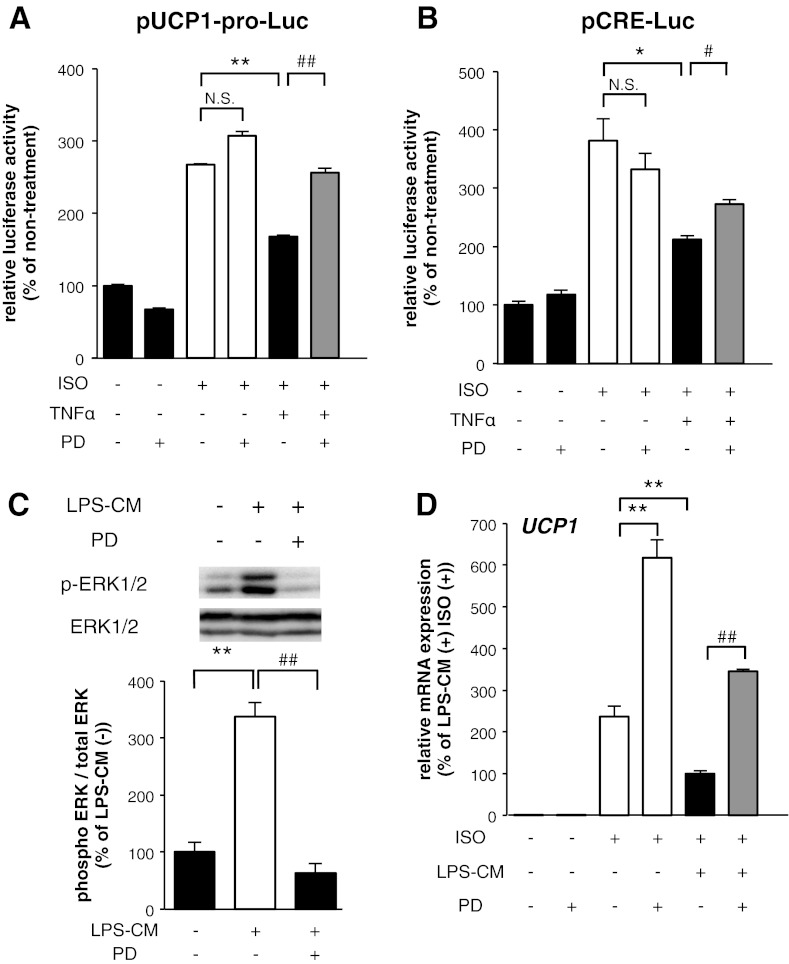
Fig. 6.
ERK activation induced by TNF-α was involved in the suppression of UCP1 promoter activation in 10T1/2 adipocytes. A and B: luciferase activity levels derived from the construct with UCP1 promoter (A) and CRE (B) in undifferentiated 10T1/2 cells incubated with TNF-α with or without PD98059 for 16 h before ISO treatment (10 μM) for 8 h. Values are means ± SE for 3–5 wells. *P < 0.05, **P < 0.01, compared with ISO-stimulated undifferentiated 10T1/2 cells. #P <0.05, ##P < 0.01, compared with ISO and TNF-α-stimulated undifferentiated 10T1/2 cells. C: 10T1/2 adipocytes were exposed to LPS-CM with PD98059 simultaneously for 15 min. The band intensities were quantified using ImageJ software. The results were calculated using data of independent experiments repeated multiple times. **P < 0.01, compared with nonstimulated 10T1/2 adipocytes. ##P < 0.01, compared with LPS-CM-stimulated 10T1/2 adipocytes. D: mRNA expression level of UCP1 in 10T1/2 adipocytes incubated with LPS-CM with or without PD98059 for 16 h before ISO treatment (10 μM) for 8 h. Values are means ± SE for 3–4 wells. **P < 0.01, compared with ISO-stimulated 10T1/2 adipocytes. ##P < 0.01, compared with LPS-CM and ISO-stimulated 10T1/2 adipocytes.

