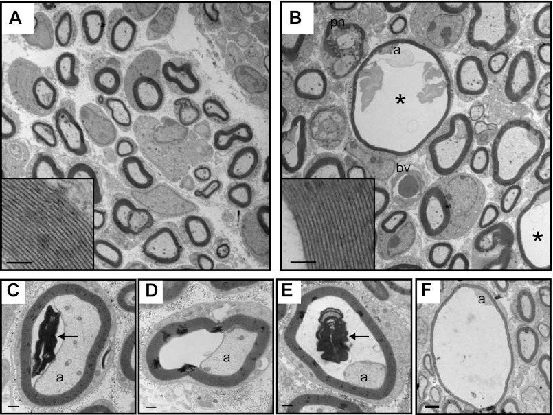
Fig. 9.
Axonal defects in KCC3-null mice. A and B: electron micrographs of P8 distal sciatic nerves isolated from wild-type (A) and KCC3-null (B) mice. Scale bar in A, 2 μm. Myelin compaction in mutant fibers (B, inset) is indistinguishable from that of wild-type (A, inset, scale bar, 500 nm). Fibers with periaxonal fluid accumulation appear only in KCC3-null nerves (asterisk, B). C–F: varying degrees of the severity of the abnormal fluid accumulation in distal KCC3-null sciatic nerve fibers. Scale bars, 500 nm. Myelin debris (arrows) is observed in the periaxonal space (C and E). a, Axon; pn, paranodes; bv, blood vessel. [Modified from Ref. 15, with permission.]

