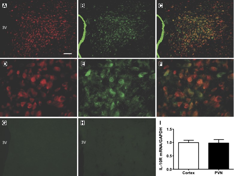
Fig. 1.
Localization of IL-10 receptor (R) on neurons in the paraventricular nucleus. A–C: low-power fluorescence micrographs (×10 magnification) showing IL-10R (green) and HuC/D (red; neuron-specific marker) immunoreactivities in the paraventricular nucleus (PVN) of a Sprague-Dawley (SD) rat. Also shown are overlays between IL-10R and NeuN. 3V, 3rd cerebroventricle; bar = 100 µm. D–F: higher power view (×40 magnification) of IL-10R and HuC/D immunoreactivities in the PVN. G and H: representative micrographs showing a lack of specific IL-10R immunoreactivity in the PVN when the primary antibody was omitted (G) or preabsorbed with an IL-10R blocking peptide (H). I: bar graph of real-time RT-PCR data showing IL-10R mRNA levels in cerebral cortex and in hypothalamic tissue containing the PVN. Immunostaining was performed on 5 SD rats. Data are means ± SE (n = 3 rats per area).

