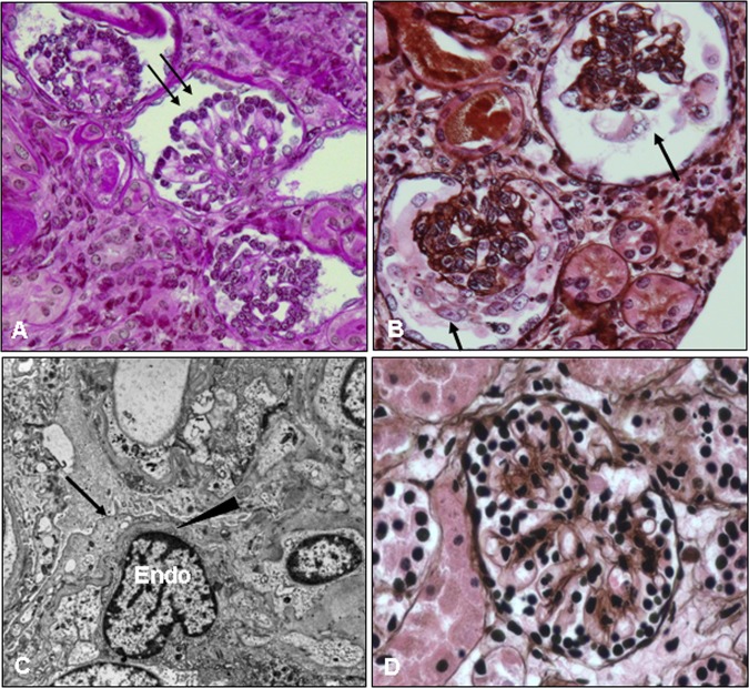
Figure 1.
Kidney biopsy of the proband at the age of 1 month. Kidney tissue was processed and stained with periodic acid Schiff (PAS) (A) and PAS methenamine (PASM) silver (B, D) for light microscopy or for electron microscopy (C). The patient's glomeruli are notably abnormal; they are small for her age and show hypercellularity, increased extracellular matrix, and contracted/collapsed glomerular tufts that are surrounded by cuboidal/immature podocytes (A, arrows) or by vacuolated/hypertrophic podocytes (B, arrows). These changes are consistent with diffuse mesangial sclerosis. A normal glomerulus from an age matched infant is shown for comparison (D). Electron micrograph images from the patient's kidney biopsy (C) show diffuse foot process effacement (arrow), thinning of the glomerular basement membrane (arrowhead), and swollen endothelial cells (Endo). This figure is only reproduced in colour in the online version.

