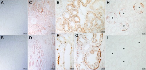
Fig. 4.

RhBG immunolabel expression in the human kidney. A and B: low-power micrograph of human kidney cortex and outer medulla, respectively, examined for RhBG immunolabel in the absence of deglycosylation. RhBG immunolabel was not detectable. C and D: low-power micrograph of human kidney cortex and outer medulla, respectively, examined for RhBG immunolabel after deglycosylation with PNGase F. Abundant RhBG immunolabel is present in a subpopulation of epithelial segments in both the cortex and outer medulla. E: high-power magnification of cortex and demonstrates strong basolateral RhBG immunolabel in the CNT. F: high-power magnification of cortical collecting duct (CCD). Abundant RhBG immunolabel is present in a subpopulation of cells. G: high-power magnification of outer medulla. Strong RhBG immunolabel is present in a subpopulation of cells in the outer medullary collecting duct (OMCD). H and I: effect of preincubation of p35 with immunizing peptide. H: no peptide was used and strong basolateral immunolabel is present. I: human kidney in which p35 was preincubated with immunizing peptide, and no significant immunolabel is observed. Antibody p35 was used for these studies. *Collecting duct lumen.
