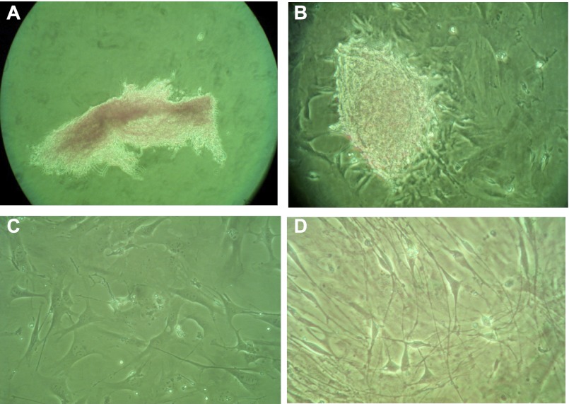
Fig. 4.
Primary cultures of esophageal stromal cells. Esophageal stromal cells were isolated using techniques previously established for colonic myofibroblast isolation, grown in primary culture and examined with an inverted microscope. On initial plating, mixed cell suspensions isolated from whole esophagus are observed partially floating and loosely attached to the plate (A). Within 24 h of plating, organoid-type structures attached to the plate bottom, with sprouting spindle-shaped cells identified (B). These sprouting cells covered the entire well within 5 days and were passaged successfully. Representative images of primary cultures between passage 5 and 15 are shown at low density (C) and when near-confluent (D).

