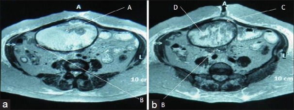
Figure 3, Case 1.

MRI abdomen-T2 W axial images of Case 1 at (a) infra-umbilical and (b) umbilical level show a large well-defined ovoid mass (A) in mid abdomen, adherent to small bowel loops (B). It has a thick hypointense capsule (C) and a central fluid filled core with whorled stripes of intermediate intensity (D).
