
Figure 5, Case 2.
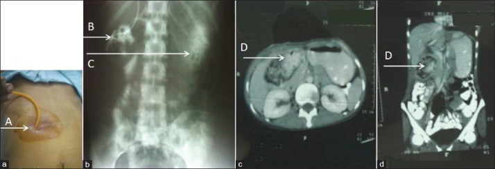
Retained sponge causing gastrocutaneous fistula. a) shows fistulogram being performed from opening at lateral end of scar b) Fistulogram performed from opening at lateral end of scar (A) shows mottled pooling of contrast in right hypochondrium (B) and contrast in stomach (C). (c) Axial CECT image at level of pylorus and (d) coronal reconstructed image through the same area show a well-circumscribed mass with contrast admixed with spongiform mottling suggestive of foreign body, in the right sub-hepatic space (D).