Figure 3.

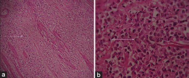
Hematoxylin and eosin stained sample (a) at ×10 and (b) at ×40 demonstrate large monomorphic tumor cells having large round cell nucleus and abundant eosinophilic cytoplasm (seen arranged in cords and nests in Figure 3a).

Hematoxylin and eosin stained sample (a) at ×10 and (b) at ×40 demonstrate large monomorphic tumor cells having large round cell nucleus and abundant eosinophilic cytoplasm (seen arranged in cords and nests in Figure 3a).