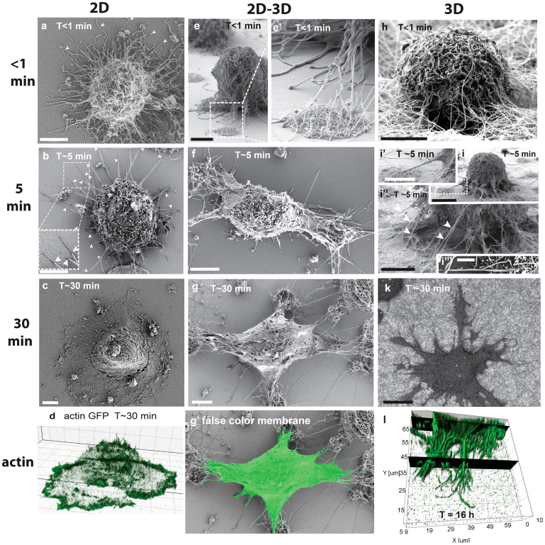
Figure 2. Time-dependent spreading events of fibroblasts on flat surfaces and in contact with NWs.
(a–d) Fibroblasts spreading on fibronectin (Fn) coated flat glass surfaces (2D). Large numbers of >10 μm long filopodia cover the entire surface of spherical cells in the first moment of substrate contact. (a arrowheads) Straight and curled filopodia are deposited randomly around the cell periphery. (b arrowheads) Within the first 5 min, most filopodia retracted and only few straight filopodia remained with distal substrate adhesions touching the surfaces under a shallow angle (kink). (c) Cells spread with a circumferential lamellipodium after passing the lag time. (d) The actin-rich lamellipodium replaced the initial surface adherent filopodia contacts (live cell imaging with actin GFP transfected cells). (e) On flat versus fibrillar interfaces, transient filopodia were initially in contact with both, flat surfaces and nanowires. (f) The transient filopodia adhered and aligned with individual nanowires and quickly directed cell spreading towards nanowire adhesion sites, while most filopodia retracted from the Fn coated flat glass surfaces. (g and Suppl. Fig. S1e) The apical membrane that is not facing nanowires was later completely depleted of filopodia, while ruffles appeared after 30 min. (g′) The false color membrane image illustrates that no lamellipodia were formed at the flat surfaces in between nanowire islands, in contrast (c, d) to flat substrates, where a full circumferential lamellipodum had occurred after the same time period. (h, i) Initial filopodia formed entangled and centripetally aligned adhesions to individual nanowires when in contact with densely NW decorated substrates. (I and Suppl. Fig. S1d) Aligned filopodia-NW adhesions guided pseudopods/membrane protrusions towards the NW anchorage site. (k) Filopodia-mediated cell spreading lead to dendritic cell shapes in the absence of typical lamellipodia-like membrane protrusions. (l) Confocal 3D reconstruction of phalloidin-alexa 546 stained actin cytoskeleton after 16 hours on 3D nanowires shows dendritic cell protrusions that display numerous filopodia, which are anchored deeply into the nanowire matrix in the absence of lamellipodia. Scale bars 5 μm with the exception of i, k = 10 μm and i ′″ = 1 μm.

