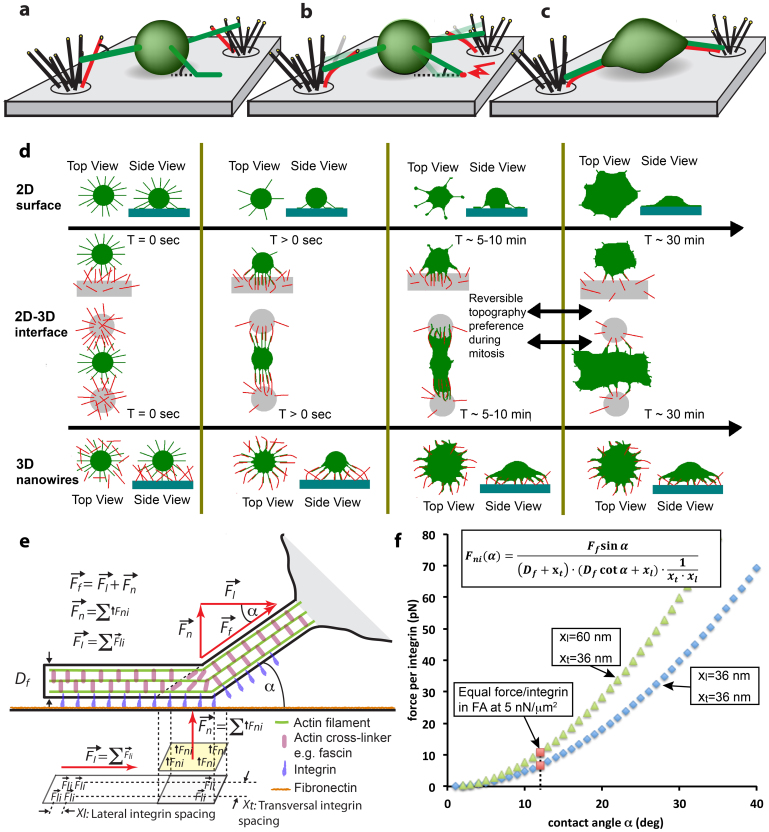
Figure 5. Underpinning mechanisms of topography sensing.
(a–c) Schematics of the topography recognizing function of filopodia. (d) Summary of our findings for the different engineered substrates and how differences in cell shape and polarity as well as the spreading dynamics are controlled by filopodia-NW interactions. (d) The first row describes for flat glass (2D) how fibroblasts undergo a rapid phase transition from filopodia-rich initial state towards a lamellipodia-mediated spreading (compare Suppl. Fig. S1b). The second row shows how cells sitting at interfaces between flat surface and NWs contact the NWs, quickly adhere, align and spread towards nanowire adhesions, while filopodia peel-off from the adjacent flat surfaces. After ~30 min, delayed lamellae/ruffle formation (Suppl. Fig. S1e) leads to migration towards the flat surface (Data not shown). This dynamic change in topography preference is reversible during mitotic rounding of fibroblasts (see Fig. 4 and Supplemental Movie M4). The bottom row shows how fibroblasts on purely nanowire decorated surfaces quickly spread via aligned filopodia-nanowire adhesions into dendritic shapes, without formation of lamellipodia. (e) Schematics showing force distributions on integrins in dependency of the angle α at which a filopodium contacts an object. (f) A simple mechanical zipping model allows to estimate the normal forces acting on individual integrins as function of the contact angle α. Since the number of integrins/μm2 within the adhesions is unknown, we calculated the normal force per integrin as function of the contact angle for two integrin densities (blue and green symbols) by assuming a filopodia traction force of 2 nN. To estimate the critical angle below which peeling will stall, we next assumed that the integrins within the filopodial shaft can sustain the same force as in focal adhesions (FAs). Cells typically apply traction forces around 5 nN/μm2 at focal adhesions (FA)63. This converts to the force per integrin as marked with red squares and thereby predicts a critical angle of close to 12° below which filopodial adhesions mature rather than peeling off before their tips detach. Supplemental figure S1c displays that only a small fraction of transient filopodia (very long ones with their base close to the flat substrate) formed angles <12°.

