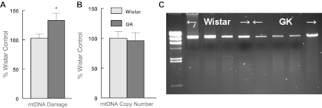
Fig. 5.

A: diabetes damages mtDNA in GK left ventricle compared with Wistar LV. mtDNA damage was assessed using the LRPCR protocol as described in methods. B: diabetes did not influence LV mitochondrial copy number. mtDNA copy number by PCR and is the ratio of mtDNA:actin DNA. Values are normalized to Wistar control and are means ± SE *P < 0.05. C: representative LRPCR mtDNA products from Wistar and GK LV.
