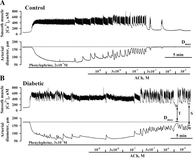
Fig. 1.
Smooth muscle cell (SMC) intracellular Ca2+ concentration ([Ca2+]i) and dilator responses of uteroplacental radial arteries to ACh are impaired in diabetic pregnancy. A and B: representative changes in SMC [Ca2+]i and diameter of arteries from control (A) and diabetic (B) rats induced by cumulative application of ACh. A: application of phenylephrine (PE) resulted in an elevation of SMC [Ca2+]i from 66 to 332 nM and in a reduction of arterial diameter from 164 to 84 μm (control). Administration of ACh reduced the SMC [Ca2+]i response and dilated the artery in a concentration-dependent manner. B: application of PE increased SMC [Ca2+]i from 101 to 401 nM and reduced the diameter of the diabetic artery from 174 to 75 μm. Dotted lines show the diameters of maximally dilated control (168 μm) and diabetic (175 μm) arteries in response to 10 μM diltiazem and 100 μM papaverine. Solid horizontal lines depict the time of exposure of arteries to PE and ACh. “T” and “S” indicate transient and sustained decreases in SMC [Ca2+]i responses and vasodilation, respectively. T is the maximal change in the ACh response calculated during 15–20 s at the end of first min of ACh application. S was calculated during the last 15–20 s of the 3-min application of each concentration of ACh. ACh-induced vasodilation was expressed as the percentage of maximal dilatation induced by papaverine and diltiazem (Dmax).

