
Fig. 4.
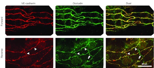
Representative segments of perfused microvessels labeled for VE-cadherin and occludin. The reverse-flow perfusions (bottom panels) showed numerous small gaps in both the occludin and VE-cadherin (arrowheads). Similar gaps were not seen in vessels perfused in the forward direction (top panels). A segment of tight junction occludin in the overlying mesothelium is visible (arrows) as a nonconnecting green shape. Scale bar, 20 μm for all images.