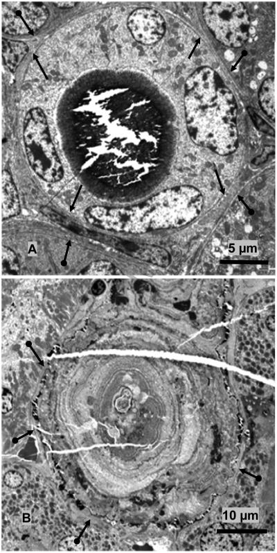
Figure 3.
TEM of Interface between crystal deposits and associated renal tissue. A. TEM of the cross section of a collecting duct. Large crystal deposit has filled the lumen lined with cuboidal cells with relatively smooth luminal plasma membrane and few cytoplasmic organelles. Both apical plasma membrane and the basement membrane (arrows) are intact. Basement membranes of the adjoining tubules are also visible (double headed arrows). B. TEM of the cross section of a large microlith completely obliterating the tubule which is surrounded by patent unobstructed tubules, with intact basement membrane 9double headed arrows). Tissue was demineralized before processing. Crystal ghosts are arranged in concentric rings (arrow heads). Center of the microlith is filled with aggregating microspheres and cellular degradation products.

