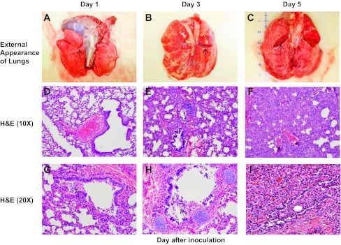
Fig. 5.

RSV infection induces pathological changes in infant baboons that resemble those in human infants with RSV infection. The external appearance of intact, fresh lung blocks recovered from RSV-infected animals on days 1, 3, and 5 following infection is shown in A–C. The cut surface (arrows) reveals the state of the lung parenchyma in each figure. Lungs demonstrate incrementally greater vascular congestion and edema over time. D–F demonstrate interstitial pneumonia with worsening inflammation, epithelial damage, and consolidation over time. The inflammatory infiltrate is more subtle on day 1 and found primarily in a peribronchiolar distribution with multifocal infiltration in the bronchiolar epithelium. Progression to day 5 reveals severe thickening of the interstitium and alveolar collapse as a result of the worsening inflammatory infiltration. H&E, hematoxylin and eosin. In G–I, there is progressive bronchiolar epithelial damage with sloughing, leading to partial to complete obstruction of some bronchioles with fragmented epithelium mixed with mononuclear inflammatory cells (arrows).
