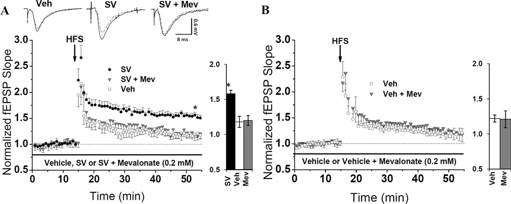
Fig. 1. Mevalonate (mev) enrichment specifically suppresses SV-mediated LTP enhancement.
(A) Hippocampal slices were pre-incubated in SV (10 µM) or veh for one hr, then moved to the recording chamber and treated with SV or Veh in the presence or absence of Mev (0.2 mM) for at least 40 min prior to HFS. The average magnitude of LTP from SV-treated slices (158 ± 0.05%, n=7 slices/7 animals) was significantly higher (* P=0.003) than that from Veh-treated slices from the same animals (118 ± 0.05%, n=7 slices/7 animals). To test if Mev enrichment can suppress this enhancement, SV + Mev-treated slices were interleaved with SV and Veh. Mev reduced (P=0.002) the LTP magnitude from 157 ± 0.04% (n=4 slices/4 animals) in SV-treated slices to 120 ± 0.07% (n=4 slices/4 animals) in slices from the same animals. (B) Mean LTP from slices pre-incubated in Veh for one hour, then treated with Veh containing either, 0.2 mM Mev or Mev's vehicle for at least 40 min pre-HFS. LTP suppression following mev enrichment is specific to SV-treated slices: LTP in Veh + Mev-treated slices averaged 121 ± 0.12% (n=6 slices/6 animals) compared to 122 ± 0.05% (n=6 slices/6 animals) LTP in Veh-treated slices (P=0.97). Waveforms show fEPSPs during baseline (dotted) and 40-min post-tetanus (solid) from slices in different treatment groups. Bar charts show LTP magnitude within each treatment group.

