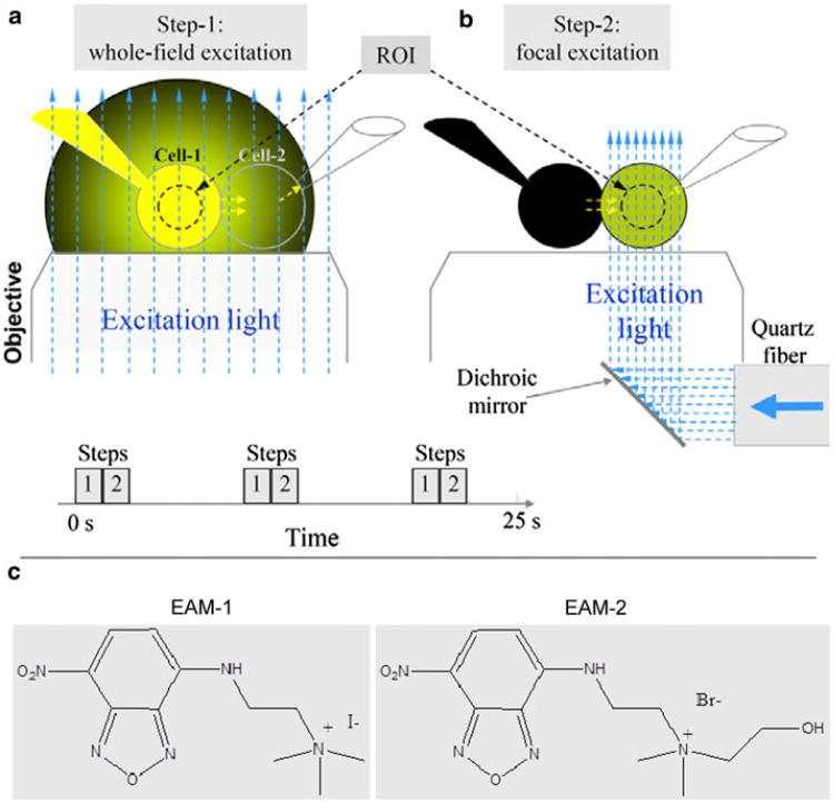
Fig. 1.
Schematics of time-lapse imaging using a dual-fluorescence excitation mode. a Whole-field excitation (vertical, blue, dashed arrows) of the cell pair with two patch pipettes. Emitted light of dye (yellow) in the pipette, and cell 1 creates a large background of scattered light around the cell pair. b The same as in a but excitation light is focused only to cell 2. This eliminates excitation of dye in cell 1 and the pipette attached to it. c Molecular formulas of positively charged fluorescent dyes, EAM-1 and EAM-2

