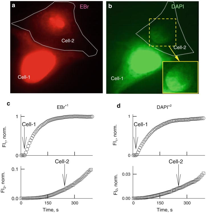
Fig. 4.
Permeability of Cx36– EGFP GJ channels to EtBr and DAPI. Representative images of HeLaCx36-EGFP cell pairs showing transfer of EtBr (a) and DAPI (b) recorded using whole-field excitation. Enhanced view of nucleus in cell 2 (inset in b) shows a gradient of fluorescence with higher intensity on the junctional side. Examples of time course of changes in EtBr (c) and DAPI (d) fluorescence in cell 1 (FI1) and cell 2 (FI2). Arrows point to the moments of patch opening in cell 1 and cell 2

