
FIG. 3.
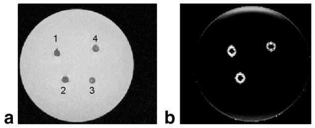
Gelatin phantom filled with different SPIO concentrations (1, 2, and 4) and agarose gel (3). a: The gradient echo image. b: A parametric map of the magnitude of the susceptibility gradient, that depicts positive contrast for the SPIO-filled holes and no contrast for the agarose. This allows a selective discrimination between SPIO and agarose, based on the induced susceptibility.