Figure 2.

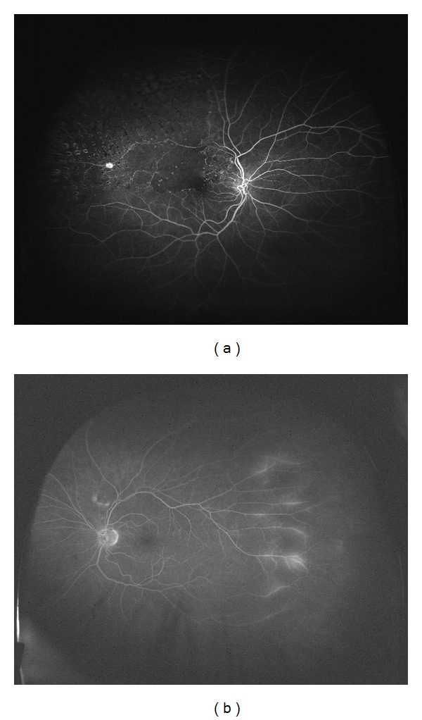
(a) Ultra wide field angiogram of a patient with branch retinal vein occlusion in the right eye. (b) The fellow eye has late peripheral leakage temporally.

(a) Ultra wide field angiogram of a patient with branch retinal vein occlusion in the right eye. (b) The fellow eye has late peripheral leakage temporally.