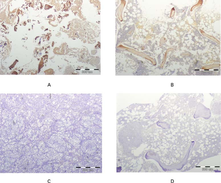
Fig. 6.
Micrographs showing immunohistochemical staining for osteocalcin (brown colour) in a) experimentally generated tissue, b) normal cancellous bone (positive control), c) kidney (negative control) and d) normal cancellous bone without antibody staining (not stained negative control). Similar positive staining is evident in the generated tissue and in normal bone sample. Negative controls show no staining to osteocalcin.

