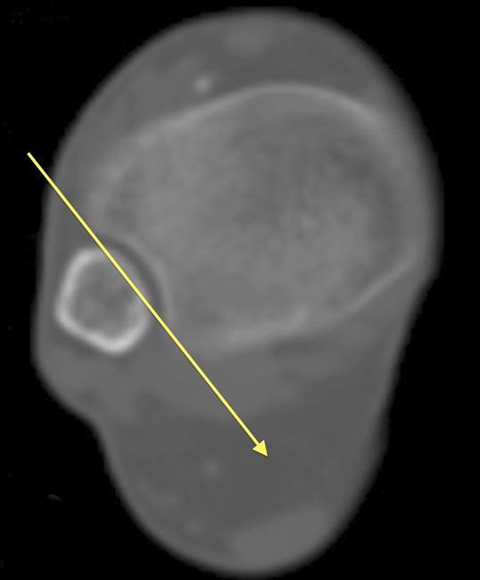
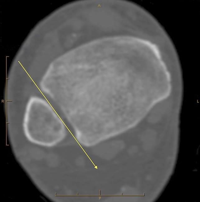
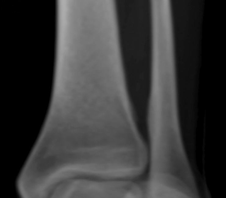
Figs. 3a - 3c.


Figures 3a and 3b – axial CT views a) in a patient with overlap between the distal tibia and fibula, showing that a plain film x-ray beam (represented by the yellow arrow) cannot be passed between the bones, and b) in a patient without overlap, with the x-ray beam (yellow line) able to pass between the tibia and fibula. Figure 3c – three-dimensional image of the patient in Figure 3b, reconstructed from CT scans. This image resembles a radiograph and can be rotated through 360° using GE Workstation software (GE Healthcare).
