Figure 2.

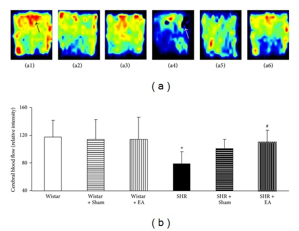
The effect of electroacupuncture on CBF in rat cerebral cortex. (a) Representative laser-Doppler perfusion images of Wistar (a1), Wistar + Sham (a2), Wistar + EA (a3), SHR (a4), SHR + Sham (a5), and SHR + EA (a6) group, respectively, acquired at week 16. Black arrow indicates the highest CBF, while white arrow indicates the lowest CBF. (b) Quantitative evaluation of CBF in six groups. Wistar: Wistar rats without any treatment. Wistar + Sham: Wistar rats with stimulation at nonacupoints. Wistar + EA: Wistar rats with stimulation at acupoints. SHR: SHR without any treatment. SHR + Sham: SHR with stimulation at nonacupoints. SHR + EA: SHR with stimulation at acupoints. Data were expressed as mean ± SD from six animals. *P < 0.05 versus Wistar group; # P < 0.05 versus SHR group.
