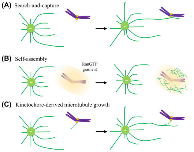
Figure 6.1. Initial interaction between kinetochores and microtubules.
(A) “Search-and-capture” model. Centrosome-nucleate microtubules undergo repeated growth and shrinkage in various directions until they are captured and stabilized by kinetochores. (B) A Ran-GTP gradient dependent “self-assembly” model. The chromatin association of the guanine nucleotide exchange factor (GEF) RCC1 produces a Ran-GTP gradient around mitotic chromosomes to simulate centrosome-independent microtubule nucleation. (C) Kinetochore-derived microtubule growth. Microtubules grow at or near kinetochore regions and later incorporate into the mitotic spindle. (For color version of this figure, the reader is referred to the online version of this book.)

